
Cartilage mechanobiology
The cartilage is a highly pressurized tissue, in which chondrocytes metabolically secrete highly charged proteoglycans that create swelling pressure in a dense and rigid collagen scaffold. The pressurization is further exacerbated by load-bearing duties which pressurize the interstitial fluids of the cartilage. As a postdoc in the Chaudhuri lab at Stanford, I am investigating how this pressurized environment influences the patho-physiology of chondrocytes especially in the context of osteoarthritis. To do this, I am studying chondrocyte behavior in 3-D in vitro models consisting of chondrocytes embedded in engineered hydrogels of tunable viscoelasticity, which differentially gates the cell-secreted nascent matrix within the hydrogel cavity, thus creating different pressurized environments [1]. I am also studying how the cellular responses to pressure are amplified by external mechanical loads. These studies elucidate new mechanisms through which cells sense the mechanical properties of their microenvironment, and highlight fundamental processes underlying the development and disease of tissues such as the cartilage.
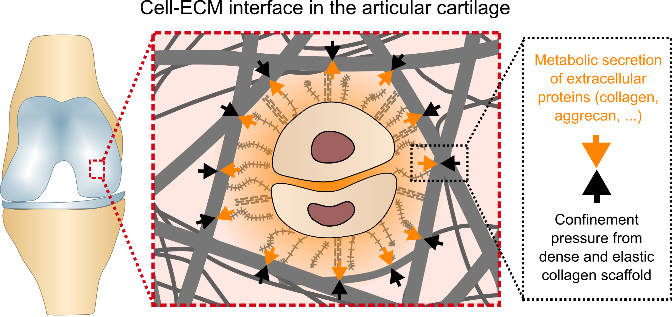
Schematic illustration of the confinement pressure generated by the cellular secretion of extracellular proteins in a highly dense and elastic collagen scaffold of the articular cartilage.
- Song, J., Jones, S., Yang, F., Bhutani, N., Levenston, M. E., & Chaudhuri, O. “Cell-Secreted Extracellular Matrix Mediates Chondrocyte Mechanosensation of Microenvironment Viscoelasticity.” Manuscript in preparation.